Aortic Dissection
What is aortic dissection? Your aorta is the main artery that carries oxygen-rich blood away from your heart to the rest of your body. The wall of your aorta is made up of three tissue layers — an inner layer (intima), middle layer (media) and outer layer (adventitia). An aortic dissection begins abruptly when a tear occurs in the inner layer of a weakened area of your aorta. Blood surges through the tear, causing the inner and middle layers to separate (“dissect”). As diverted blood flows between the tissue layers, the normal blood flow to parts of your body may be slowed or stopped, or the aorta may rupture completely. Aortic dissection is a life-threatening condition that can cause sudden death if it is not recognized and quickly treated.
Where is the aorta? The aorta runs throughout your torso. It begins at the main pumping chamber of your heart (the left ventricle), extends up through the front middle of your chest, arches from front to back under the base of your neck, then travels downward along the front of your spine — through your chest (thoracic aorta) and abdomen (abdominal aorta) — before branching just below your navel to two other arteries called the right and left common iliac arteries.
Are there different types of aortic dissection? There are two main types: Stanford Type A Aortic Dissection: This type of dissection occurs in the first part of the aorta, closer to the heart, and can be immediately life-threatening. It usually requires emergency open chest surgery to repair or replace the first segment of the aorta where the tear started (ascending aorta +/- the arch and/or aortic valve). This is a more common type of dissection than Type B, and the dissection of the aorta usually extends through the entire length of the aorta. Stanford Type B Aortic Dissection: This type of tear begins farther down the aorta (descending aorta beyond the arch), and farther from the heart. Like the type A dissection, this usually extends from the descending aorta into the abdominal segment (abdominal aorta), but doesn’t involve the first part of the aorta in the front of the chest. Surgery may or may not be needed immediately, depending on exactly where the dissection is located and if it is or isn’t cutting off blood flow to your organs. These operations usually can be performed with a stent-graft device inserted into the aorta. Another classification system (DeBakey Classification) defines dissection by three types. Type 1 originates in the ascending aorta and extends through the downstream aorta. Type 2 originates and is limited to the ascending aorta (both would be considered Stanford Type A). Type 3 originates in the descending aorta and extends downward (similar to Type B).
What’s the difference between aortic
aneurysm, aortic rupture and aortic dissection?
An aortic aneurysm is a bulge — like
a bubble or a balloon — in a weakened area of the wall of the aorta or across
an entire segment of the aorta. Aortic aneurysm can lead to aortic rupture and
aortic dissection.
An aortic rupture is a complete tear
through all three layers of the aorta — like a rip or a hole — in the wall of
the aorta. Blood bursts through the hole into the surrounding body cavity.
An aortic dissection is a tear in
the inner aortic layer that allows blood to enter and further separate the inner
and middle layers of the wall of the aorta and typically extends over a long
length of the aorta in either direction and may extend into branch vessels
originating from the aorta.
What are the signs and symptoms of
aortic dissection?
The most common characteristic of
aortic dissection is its abrupt start. It can happen at any time, while doing
anything, or at rest or when you’re sleeping.
Common signs and symptoms include:
- Sudden severe, sharp pain in your
chest or upper back; also described as a tearing, stabbing or ripping feeling.
- Shortness of breath.
- Fainting or dizziness.
- Low blood pressure; high suspicion
when there’s a 20 mmHg pressure difference between arms.
- Diastolic heart murmur, muffled
heart sounds.
- Rapid weak pulse.
- Heavy sweating.
- Confusion.
- Loss of vision.
- Stroke symptoms, including weakness
or paralysis on one side of your body, trouble talking.
What causes aortic dissection?
Aortic dissection happens because
there is an underlying, slow breakdown of the cells that make up the walls of
your aorta. The breakdown has likely been going on silently for many years
before the weakened area of the aortic wall finally gives way, resulting in a
tear, which leads to the aortic dissection.
Why does the aortic wall weaken in
some people and not others? It’s believed that most aortic dissections are
caused by an underlying vulnerability that may be inherited. In others, the
stress to the aortic wall from constant high blood pressure can weaken the
aorta wall in susceptible people, resulting in a tear and dissection.
Aortic dissection in the ascending
aorta (the section closest to the heart where the pressure is the highest) is
nearly two times more common than those that occur in the descending aorta.
Tears in the aorta typically occur in areas where the stress on the wall of the
aorta is highest.
What factors can increase the risk
of developing aortic dissection?
Factors that can increase your risk
for developing aortic dissection include:
- Ongoing high blood pressure
(hypertension). This is the most important risk factor. High blood pressure
causes direct damage to the layers of aortic tissue, causing loss of elastic
fibers, breakdown of the wall structure and increased wall stiffness.
- Atherosclerosis (or buildup of
plaque in the arteries)/high cholesterol and smoking.
- Aortic aneurysm. This is an abnormal
enlargement or bulge in the aortic wall.
- Aortic valve disease.
- Congenital (“born with”) heart
conditions like a bicuspid aortic valve (has two leaflets instead of the normal
three) or Turner syndrome.
- Connective tissue disorders, such as
Marfan syndrome and Ehlers-Danlos syndrome. These are genetically linked
problems that can be passed down to family members.
- Other hereditary thoracic aortic
conditions that primarily affect the aorta that are also genetically caused.
- Family history of aortic dissection.
- Vasculitis, specifically aortitis.
This inflammatory disease affects the body’s blood vessels.
- Traumatic injury to the chest (e.g.,
after a high-speed car crash or serious fall from a height of> 20 feet).
- Age between 50 and 65 years. The
aortic wall loses its elasticity with age.
- Being pregnant and having high blood
pressure during delivery.
- Activities that extend periods of
high blood pressure, such as cocaine or amphetamine use.
- Strenuous powerlifting may increase
the speed of development of aneurysms or dissection in susceptible people.
What complications can result from
aortic dissection?
Aortic dissection can lead to:
- Stroke.
- Aortic valve damage.
- Damage to internal organs.
- Fluid buildup between the heart
muscle itself and the sac covering the heart. This condition, called cardiac
tamponade, puts pressure on the heart and prevents it from working properly.
- Death.
How is aortic dissection diagnosed?
Aortic dissection must be diagnosed
quickly, in case immediate surgery is needed. The healthcare team needs to
determine if you have aortic dissection or other health conditions, such as
heart attack and stroke, which produce similar symptoms. Tests that may be
ordered include:
Chest X-ray: This test uses a small
amount of radiation to create an image of the structures within your chest,
including your heart, lungs, blood vessels (including the aorta) and bones.
This test is not very specific but is quick and may direct the diagnosis.
Computed tomography (CT) scan: This
test provides the best view of the aorta during an emergency and can be
performed rather quickly to look for aneurysm or dissection. For aortic
imaging, intravenous (IV) contrast may be needed.
Transthoracic echocardiogram: This
test uses ultrasound to provide moving pictures of your heart valves and
chambers and the first portion of the aorta (the aortic root).
Transesophageal echocardiogram
(TEE): This test shows more detailed pictures of your heart valves and chambers
than a transthoracic echocardiogram and better views of your thoracic aorta.
The ultrasound probe is placed through your mouth into your esophagus, which
runs directly behind your heart and in from of your descending aorta.
Magnetic resonance imaging (MRI):
This test uses a large magnet and radio waves to produce detailed images of
your organs and the structures inside your body, including your aorta. It
provides moving pictures of your heart valves and chambers and blood flow
through your aorta. This test may take more time to perform than a typical CT
scan and so is less often used in emergencies.
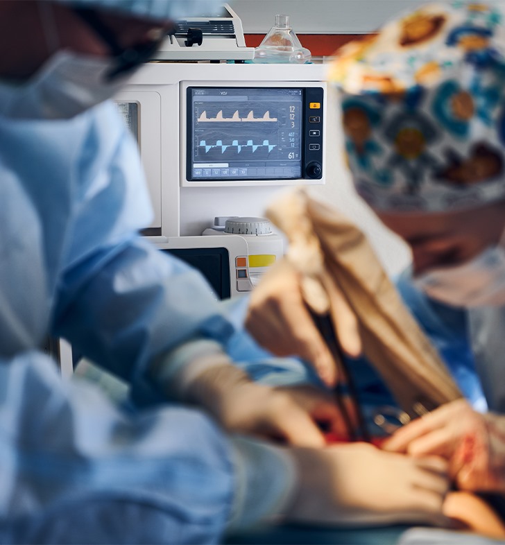
How is aortic dissection treated?
Treatment of aortic dissection
depends upon the location of the tear and dissection. Immediate surgery is
needed for Type A aortic dissection (i.e., when it involves the first part of
the aorta close to the heart). Type B aortic dissection requires emergency
surgery if the dissection cuts off blood flow to your vital organs including
your kidneys, intestines, legs or even your spinal cord. Urgent surgery is
needed if there are certain high-risk features noted on CT scan imaging. Less
severe cases may be treated with medication initially, delaying surgery until
complications develop.
Surgery and Endovascular Treatment
Surgical options include:
Graft replacement: With this
approach, a portion of the damaged section of the aorta is removed and a
synthetic fabric tube (graft) is sewn directly in its place.
Endovascular stent-graft repair:
With this approach, a stent graft — a synthetic fabric tube supported by metal
wire stents (like a scaffold) — is used to repair the aorta from within.
Endovascular surgery involves making the repair inside your aorta. A small
incision is made in your groin and a catheter, with the fabric-lined stent
attached, is delivered and deployed into the aorta under x-ray guidance. At the
repair site, the stent graft is released and — like a spring or umbrella —
opens up, relining and providing reinforcement to the weak area in the aorta.
Hybrid approach: With this approach,
a combination of conventional open surgery and endovascular stent-graft
technique is used to repair the aorta. This is used when the repair must extend
into the aortic arch where branch vessels to the brain and arms arise. This may
be performed during the emergency operation for Type A dissection or as a
two-stage repair with a bypass from a vessel in the neck to help set up an
endovascular repair for Type B dissection. One of the most common hybrid
procedures is called the “elephant trunk” or “frozen elephant trunk” procedure.
First, the segment of the aorta close to the heart as well as the aortic arch
(the segment of the aorta that supplies blood to the brain) is replaced and
repaired. An additional graft, or stent graft, is left hanging into the
descending aorta, like the trunk of an elephant. The graft is ready to receive
the endovascularly placed stent-graft when the second surgery is performed.
Medications
Medications, such as beta blockers,
may be prescribed to lower heart rate and blood pressure. In some Type B aortic
dissection cases, medication alone may be used to treat the dissection
initially. Surgery may be able to be delayed for months to years depending on
the severity of the tear and extent of dissection.